Innovation in Medical and Drug Discovery Visualization Equipment through the Fusion of Electrical Tomography and Machine Learning

-
- Principal Investigator
Professor / Masahiro TAKEI
- Affiliation
Graduate School of Engineering, Chiba University
Researchmap
ORCID ID
- Principal Investigator
Early diagnosis is critical to the treatment of diseases. Several patients worldwide suffer from various diseases. Such patients include not only those treated in hospitals but also those treated at home. However, extensive and expensive visualization equipment such as medical X-ray computed tomography (CT) and magnetic resonance imaging cannot be operated in common households. Therefore, it is difficult to detect the onset and progression of diseases at an early stage. To address this issue, there is an urgent need for a simplified biological tissue visualization technology that allows disease diagnosis at home.
Our research group is currently working towards the development of a visualization technology using electrical tomography. Electrical tomography, also known as electrical CT, involves placing a multi-electrode sensor around the human body and measures the electrical resistance and capacitance between several electrode pairs. Thereafter, the conductivity and permittivity distributions of the cross-sections of the human body are reconstructed. Compared with X-ray CT, electrical tomography is far superior in terms of safety and cost. Moreover, the equipment can be easily operated by anyone, similar to a body analyzer.
Through this study, we aim to develop an innovative technology based on electrical tomography that can visualize the cross-section of the human body in a four-dimensional (4D) space (3D space + 1D time). In addition, our goal is to build a next-generation medical setup by developing a system that allows doctors to perform remote diagnoses based on 4D images, allowing patients to monitor their health status on a daily basis.
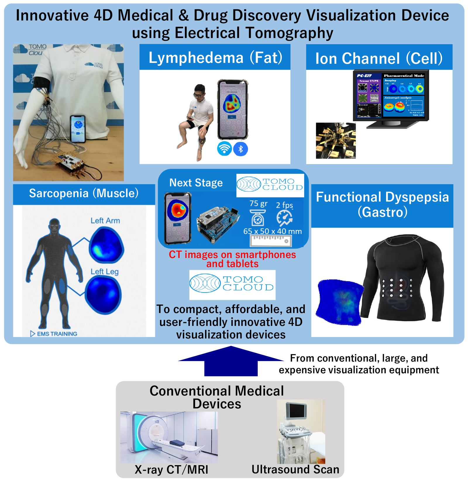
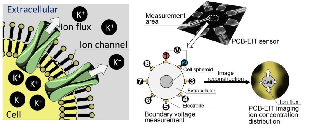
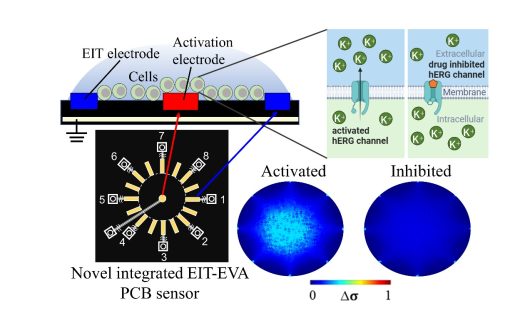
We are currently promoting a wide range of studies in the fields of medicine, nursing, and drug development. For example, in the case of lymphedema, it is crucial to test the condition of fat tissue because it is related to the condition of the adipose tissue and the flow of lymphatic fluid. Our technology demonstrates an ability to render fat visible. Similarly, it enables visualization of muscles in sarcopenia, a condition wherein muscle strength decreases due to aging or a disease. In functional dyspepsia, we can visualize the stomach when symptoms such as stomach pain or fatigue are prevalent, but the cause of discomfort, including diseases such as gastric cancer or stomach ulcer, cannot be accurately determined. In addition, cells such as ion channels (membrane proteins) can also be visualized. We support research that demonstrates broad applicability in medicine, nursing, and drug discovery.
Furthermore, we aim to establish a Chiba University venture company. The venture company will work on manufacturing and selling devices for medical and drug research based on the innovative 4D visualization technology and create societal awareness to enable implementation of the breakthrough technology.

