
MULTIMODAL MEDICAL ENGINEERING
TOWARD IMAGE-BASED DISEASE DIAGNOSTICS
-Novel, high-precision imaging techniques are being used to provide non-invasive disease diagnosis and guide future treatments.
Research Keywords: Medical Engineering, Physical Measurement,Multimodality
Non-invasive, precise diagnosis of diseases using advanced medical imaging technologies could one day replace traditional techniques based on biopsy and dissection. With this prospect in mind, researchers at the Multimodal Medical Engineering program are combining medical and engineering expertise to create novel, non-invasive imaging-based methods for diagnosing and treating disease.
The program uses various scanning techniques, or ‘modalities’, including computerized tomography, magnetic resonance imaging (MRI) and ultrasound, to analyze cellular and organ variations caused by different diseases.
The program’s goal is to develop highly precise, non-invasive imaging methods, while boosting existing imaging techniques to improve resolution and enable more-detailed tissue analysis, says program leader Hideaki Haneishi.
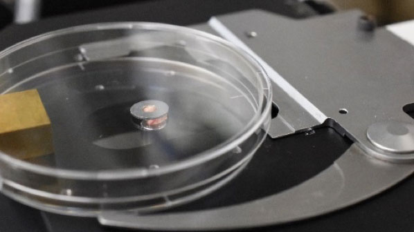
“Many current disease diagnostics, particularly for cancer, involve highly invasive, unpleasant procedures to clarify the type and stage of the disease,” explains Haneishi. “We are investigating, for example, the development of a system to diagnose breast cancer using very-high-frequency ultrasound.”
Rather than physically sampling and dissecting lymph nodes from potentially cancerous tissue, his team has shown that it is possible to analyse cell characteristics of lymph nodes using ultrasound. Their technique highlights metastasis positive and negative cells in lymph nodes, enabling rapid, painless diagnosis.
Haneishi and his team are also devising ways to combine data from two or more modalities. For example, in 2016 they developed an image registration method that transforms two data sets into one coordinated system using MRI and pathological images of brain tumors.
“If we can establish the technology for registering two kinds of images, then MRI features that are strongly related to the pathological features of tumors can be highlighted. This may eventually allow us to diagnose using only MRI,” says Haneishi. “Determining the relationships between different data sets helps us build up detailed pictures of what is going on inside the body, not only for diagnosis but also to improve our understanding of disease development,” he adds.
NOVEL OPTICAL PROBES
Alongside conventional imaging techniques, the researchers are also exploring novel optical probes. One example involves a method for monitoring microcirculation — the flow of red blood cells — in the body. The novel probe comprises multicolored light-emitting diodes (LEDs). As the LED light is scattered through the tissue, it is partly absorbed by molecules in the red blood cells, generating a clear, detailed picture of the cells and their microcirculatory network.
Building on previous work, the team hopes to use this probe to estimate features such as the oxygen saturation of each blood vessel in areas around suspected tumors in real time. This could aid diagnosis and further our understanding of vascular behavior in and around tumors.“Ultimately, we would like to build on these imaging techniques and use machine learning and artificial intelligence technologies to relate measured signals and diagnoses, to develop a comprehensive understanding of different diseases,” says Haneishi.
The Multimodal Medical Engineering program has access to highly specialized equipment and technologies that can help realize researchers’ ideas.
“We welcome engineers and medical researchers who wish to work at the cutting-edge of research and development with direct clinical applications,” says Haneishi.
(CHIBA RESEARCH 2020)Members
Principal Investigator
| Name | Title, Affiliation | Research Themes |
|---|---|---|
| HANEISHI Hideaki | Professor, Center for Frontier Medical Engineering | Medical Image |
Co-Investigatior
| Name | Title, Affiliation | Research Themes |
|---|---|---|
| YAMAGUCHI Tadashi | Professor, Center for Frontier Medical Engineering | Ultrasonic Measurement |
| HAYASHI Hideki | Professor, Center for Frontier Medical Engineering | Digestive Surgery |
| YOSHIDA Kenji | Associate Professor, Center for Frontier Medical Engineering | Ultrasonic Measurement |
| SAITO Kazuyuki | Associate Professor, Center for Frontier Medical Engineering | Electrical Engineering |
| KAWAMURA Kazuya | Assistant Professor, Center for Frontier Medical Engineering | Medical Robotics |
| UNO Takashi | Professor, Graduate School of Medicine | Clinical Imaging |
| NAKAGUCHI Toshiya | Professor | Center for Frontier Medical Engineering, Medical Image Processing, Color Dynamics |
| SUGA Mikio | Associate Professor, Center for Frontier Medical Engineering | MRI Engineering |
| IKEHARA Yuzuru | Professor, Graduate School of Medicine | Experimental Pathology, Sugar Chain Biomarker |
| IWADATE Yasuo | Professor, Graduate School of Medicine | Neurosurgery, Neuro-Oncology |
| HAYANO Koichi | Associate Professor, Graduate School of Medicine | Surgery, Sugar Chain Biomarker |
| LIU Hao | Professor, Graduate School of Engineering | Biomechanics, Biomimetics |
| TSUBOTA Ken-ichi | Professor, Graduate School of Engineering | Biomechanics |
| YU WenWei | Professor, Center for Frontier Medical Engineering | Robotics, Machine Learning, Object Recognition |
| SUYARI Hiroki | Professor, Graduate School of Engineering | Information Engineering |